Profs. Hengyong Yu and Zeinab Hajjarian’s Work Is Supported by NIH

09/01/2024
By Edwin L. Aguirre
Advances in medical imaging technologies have revolutionized how diseases and physical injuries are diagnosed, monitored and treated, leading to better patient outcomes and more personalized care.
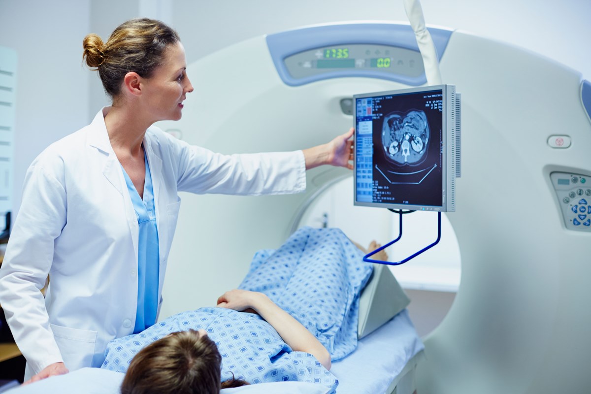
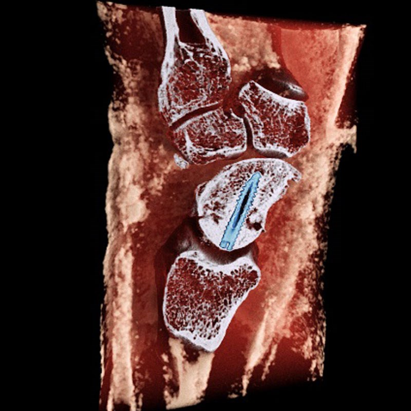
Modern high-resolution imaging techniques, like computed tomography (CT) scans, provide detailed images of the body’s internal structures, allowing for the early detection and diagnosis of diseases such as cancer and internal injuries such as fractured bones. Image-guided surgical technologies allow surgeons to perform minimally invasive procedures with high precision, thereby reducing recovery time, minimizing scarring and lowering the risk of complications.
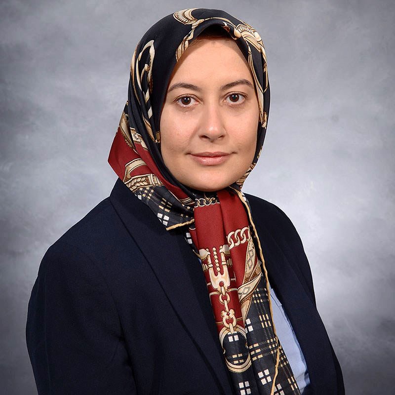
Two UML faculty researchers—Prof. Hengyong Yu of the Department of Electrical and Computer Engineering and Asst. Prof. Zeinab Hajjarian of the Department of Biomedical Engineering—are spearheading efforts to improve these vital imaging tools. Their projects are supported by grants totaling $5.1 million from the National Institutes of Health’s National Institute of Biomedical Imaging and Bioengineering.

Yu is developing technology that would greatly improve cardiac CT scans, which doctors currently use to diagnose cardiovascular diseases so that timely, life-saving treatment and preventive measures can be implemented. He is also helping to improve the image quality and resolution of photon-counting CT scans.
Hajjarian’s project aims to develop advanced optical imaging and sensing technology using laser and high-speed cameras to enhance our fundamental understanding of diseases and their progression, particularly breast cancer.
The researchers hope their efforts will contribute to more accurate prognoses and ultimately improve patient outcomes and survival rates.
'Freezing' the Beating of the Heart
Heart disease is the leading cause of death for men and women in America, according to the U.S. Centers for Disease Control and Prevention (CDC).
One person dies every 34 seconds in the United States from cardiovascular disease, the CDC reports, and this costs the country about $229 billion each year in health care services, medicines and lost productivity.
A cardiac CT scan, or coronary CT angiography, is an imaging procedure widely used in hospitals that combines a series of X-ray images and processes them on a computer to create detailed, cross-sectional views of the patient’s heart and blood vessels. Physicians use them to check for narrowed or blocked arteries and other serious heart conditions. The biggest drawback with this imaging technique is that the movement of the beating heart can result in blurry images.
Yu is leading a team of researchers from UMass Lowell, Rensselaer Polytechnic Institute and Vanderbilt University Medical Center working to develop a new image-reconstruction algorithm based on artificial intelligence to effectively “freeze” the beating heart in CT images. The project is supported by a four-year grant worth more than $2.4 million from NIH.

“Our algorithm would eliminate the blurring movement of the coronary arteries in X-ray images and help doctors analyze plaque buildup on the walls of the arteries, which is the main cause of heart attacks,” Yu says.
“Moreover, our method will not require patients to hold their breath during the CT exam and will eliminate the need to use beta-blocker drugs to slow down the patients’ heart rate,” he says.
According to Yu, the team’s AI-based computational framework would radically improve the image quality of existing CT scanners and would benefit patients who suffer from conditions such as tachycardia (rapid heartbeat) and arrhythmia (irregular heartbeat) that commonly occur in older adults, many of whom experience atrial fibrillation (rapid, irregular heart rhythm).
“Our project will combine two innovative image-processing algorithms—compressed sensing and deep learning—to reconstruct cardiac CT images at very high resolution and with lower radiation exposure to patients compared with traditional CT scans,” Yu notes.
He says their technique could allow them to help build powerful, low-cost cardiac CT scanners and possibly to retrofit older models to perform cardiac CT exams, dramatically expanding the capability of these systems and making higher-quality cardiac CT scans available in many underserved communities worldwide.
The Power of CT Image Reconstruction
Yu’s second award—a four-year, $2.3 million NIH grant—aims to enhance photon-counting CT scans by using the power of AI technology for 3D color CT imaging.
“This technique fully utilizes the energy spectrum of the X-ray source and the energy-discriminating capability of the photon-counting detector,” says Yu, who is the project’s principal investigator. “This novel technology generates multi-energy images at high spatial resolution, so it outperforms conventional CT imaging and contrast agents as well as pharmaceutical drugs in characterizing soft tissues.”

Each year, more than 80 million CT scans are performed in the United States. According to Yu, photon-counting CT offers not only fast, noise-free imaging, but also a lower dose of ionizing radiation compared with traditional CT scanners.
“This opens a new door to huge opportunities for imaging at the functional, cellular and even molecular level, using novel contrast agents such as gold and bismuth nanoparticles,” he says.
Yu’s goal is to develop deep-learning algorithms and artificial neural networks that can automatically extract and analyze information or find hidden patterns from large amounts of data to reconstruct the images.
Yu’s co-investigators are Profs. Yan Luo (electrical and computer engineering) and Yu Cao (computer science). Doctoral students Shuo Han and Bahareh Morovati are assisting Yu in the lab work. External collaborators include researchers from MARS Bioimaging Ltd., a medical imaging company based in Christchurch, New Zealand, and Rensselaer Polytechnic Institute.
Improving Breast Cancer Imaging
Breast cancer is the most commonly diagnosed cancer and the second most common cause of cancer-related death in women worldwide, according to the CDC.

Increased awareness and screening have led to the detection of early-stage small lesions. Accurately delineating the malignant tumor during surgical removal is likewise important so that as much breast tissue as possible can be preserved. But this can be a challenge, especially to those with dense breast tissues.
It is estimated that more than 15% of women who undergo breast conservation surgery have to return for additional surgery due to the inadequate removal of the malignant tumor and the risk of local recurrence.
“This creates a significant physical and mental burden on the patients and health care resources,” says Hajjarian, who joined UMass Lowell from the Massachusetts General Hospital’s Wellman Center for Photomedicine in 2023.
According to Hajjarian, her goal is to develop an innovative optical imaging technique, called laser speckle rheological microscopy, that would allow for rapid, noncontact high-resolution mapping of the viscoelastic properties within the malignant tissue. She says that by identifying the micromechanical features that are shared between invasive diseases, this approach would help differentiate the tumor from the surrounding healthy tissues.
“Light reflected from the tumor could capture information on its mechanical properties, such as stiffness,” she explains. “Our goal is to optimize the resolution of our laser technique to reduce imaging time and identify specific mechanical features that indicate invasive behavior.”
The project is funded with a three-year, $400,000 grant from NIH, with Hajjarian as principal investigator. Assisting her with the lab research were Alyre Blazon-Brown, a UML master’s student in physics, and Kayma Konecny, an undergraduate student from the University of Arizona’s Wyant College of Optical Sciences in Tucson.
