Project Is Supported with $960,000 in Grants from Massachusetts Life Sciences Center and MARS Bioimaging

UMass researchers are developing contrast agents that are specifically designed to recognize breast cancer cells and bind to them, thereby amplifying the X-ray signal for tumors and enhancing their visibility in spectral CT scans.
02/03/2021
By Edwin L. Aguirre
A team of researchers led by Chemistry Asst. Prof. Manos Gkikas is developing an advanced X-ray imaging method that aims to improve the detection and diagnosis of breast cancer.
The noninvasive technology uses dyes, called contrast agents, that are specifically designed to recognize molecularly breast cancer cells and bind to them. The dyes will amplify the X-ray signal for tumors when imaged with a special, state-of-the-art computed tomography (CT) scanner, called “photon-counting spectral CT.”
“The contrast agents, combined with spectral CT and machine learning, could lead to a more precise diagnosis of the disease and assist significantly in early intervention,” says Gkikas, who is the principal investigator (PI) for the project.
Other team members include Prof. Hengyong Yu of the Department of Electrical and Computer Engineering and Assoc. Prof. Mary Rusckowski of the UMass Medical School Department of Radiology as co-PIs, and MARS Bioimaging Ltd. as industry partner.
“To the best of our knowledge, this is the first time this type of combined research is being done,” says Gkikas. “It highlights the importance of X-ray CT in medicine, a field where UMass is strong.”
The project is funded with a three-year, $750,000 grant from the Massachusetts Life Sciences Center, with UMass Lowell getting $590,000 and the rest going to UMass Medical School. In addition, MARS Bioimaging, a New Zealand-based company that is a pioneer in photon-counting spectral CT, will sponsor a postdoctoral researcher to work at UMass Lowell for three years, for a total of $210,000.
“The majority of the Mass Life Sciences Center funding will be used to purchase a photon-counting spectral CT scanner from MARS Bioimaging,” says Gkikas.
A Silent Killer
Breast cancer is the most common type of cancer in American women and the second leading cause of cancer deaths among women, according to the American Cancer Society. The organization estimates that in 2021, about 281,550 new cases of invasive breast cancer will be diagnosed in women in the United States, and an estimated 43,600 patients will die from the disease.

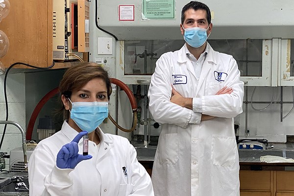
Chemistry Asst. Prof. Manos Gkikas, right, with Ph.D. student Shayesteh Tafazoli, who is holding a vial of gold-based nanoparticle contrast agent for use in X-ray spectral CT.
Unlike images from conventional CT scanners, the multicolor, 3D X-ray images generated by spectral CT can help visualize tissue composition in the body based on the density and the atomic number of chemical elements found in those tissues.
“This can assist radiologists in discriminating between healthy and cancerous tissues in the body,” explains Yu. “They can use those images to determine if a cyst or a tumor requires further examination or biopsy.”
“Unlike other medical imaging techniques such as PET and SPECT, CT uses lower energy for imaging and is faster, safer and more comfortable for patients,” says Gkikas.
Gkikas notes, however, that the widely used iodine-based contrast agents approved by the U.S. Food and Drug Administration allow only for fast screening lasting several minutes before they are excreted from the body, while other metal-based contrast media reported in preclinical studies lack the ability to specifically target cancer cells.
“In our approach, we are designing metal-based nanomaterial contrast agents that could stay in the body for a prolonged period due to their high specificity for tumors,” Gkikas says. “They can accumulate at the cancer site, based on what breast cancer cells produce or feed from, and enhance the CT signal to better visualize the tumor.”
“The resulting data can then be amplified even further using image reconstruction algorithms and machine learning, enabling us to track a tumor’s progression in primary breast cancer,” adds Yu.
According to Gkikas, if the technology is successful, it could be expanded later to detect secondary metastatic cancers – those that usually emerge 4 to 10 years after treatment of the primary cancers and have spread to other tissues and organs. It could even be used to improve early diagnosis of other diseases.
“We believe our methodology can provide significant improvement in detecting breast cancer, arthritis and other diseases, including COVID-19,” says Gkikas.
Last summer, Gkikas was awarded a $9,000 seed grant by the university’s Office of Research and Innovation to apply the imaging technique to detect COVID-19 and track the progress of lung inflammation, from mild symptoms to severe illness. That project is ongoing.
In addition to the faculty researchers, Ph.D. student Shayesteh Tafazoli is working in Gkikas’ lab on preparing disease-targeting CT contrast agents, as well as testing their cell biocompatibility, while Ph.D. students Dayang Wang and Yongshun Xu are working in Yu’s lab on machine learning for X-ray image reconstruction and analysis.
